3D-DOCTOR Display Functions
3D-DOCTOR displays 2D and 3D images in several ways for the best visualization quality and the easiest navigation between slices. 3D-DOCTOR not only supports most commonly used image types, including 8 or 16-bit grayscale and color images, but also display 3D point cloud data as a surface model or volume image.
With 3D-DOCTOR, you can:
- Display cross-section CT/MRI images in single slice mode and montage mode to see all slices at the same time.
- Separate the slices from a scanned film and then perform 3D imaging and visualization applications.
- Use the animation function to fly through all slices and create a movie.
- Make on-screen measurements for area, length, and pixel density using just the mouse.
- Add annotations and markers on the image.
- Reslice a volume image to see the profile.
- Enhance the image display with one of the many advanced image processing functions.
- Create 3D surface models using surface rendering and calculate 3D volume and surface area.
- Create 3D volume display using volume rendering and look at the image from any angle.
- Display your image in pseudo color or simply create your own color palette to show pixels you are interested.
- Click here to see sample displays.
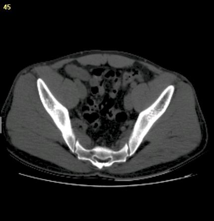
Step 1. Original CT image | 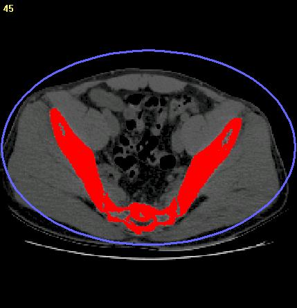
Step 2. Segmentation | 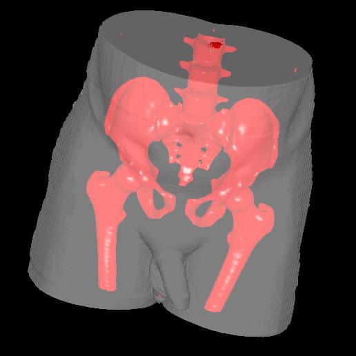
Step 3. 3D mesh model created |
3D-DOCTOR is an advanced 3D modeling,
image processing and measurement software for MRI, CT, PET, microscopy, scientific, and industrial
imaging
applications. 3D-DOCTOR supports both grayscale and color images stored
in DICOM, TIFF, Interfile, GIF, JPEG, PNG, BMP, PGM, RAW or other image file
formats. 3D-DOCTOR creates 3D surface models and
volume rendering from 2D cross-section images in real
time on your PC. The following rendering is created from a CT scan of a mummy using
3D-DOCTOR: You can export the mesh models to STL (ASCII or Binary), DXF, IGES, 3DS,
OBJ, VRML, PLY, XYZ and other formats for surgical
planning, simulation, quantitative analysis and rapid prototyping
applications. You can calculate 3D volume and make other 3D measurements for quantitative analysis.
3D-DOCTOR's vector-based tools support easy image data
handling, measurement, and analysis. 3D CT/MRI images can
be re-sliced easily along an arbitrary axis. Multi-modality images can
be registered to create image fusions. Misaligned slices can be
automatically or semi-automatically aligned using 3D-DOCTOR's image
alignment functions. The 3DBasic
scripting tool makes it easy to create Basic-like
sophisticated 3D imaging programs. Get 3D-DOCTOR today
and visualize your images in 3D. 3D-DOCTOR
is approved by FDA (US Food and Drug Administration 510K clearance) for
medical imaging and 3D visualization applications. It has been named the Top 3D Imaging Software by Scientific Computing
& Instrumentation Magazine in the Year 2002 and Year 2000 Annual Technology Leaders Issue. 3D-DOCTOR is currently being used by leading hospitals,
medical schools and research organizations around the world.